
 العربية
 Español
 中文
中文 Deutsch
 Français
 Português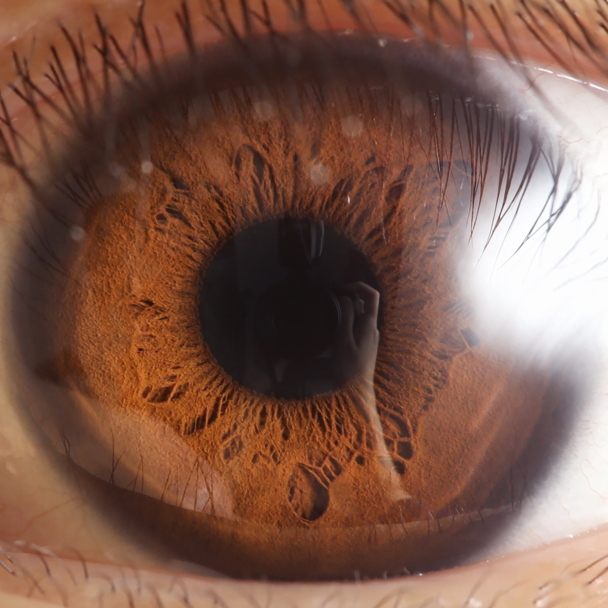
The center of your vision - the part you are using to read this sentence - is built by cells that change their minds. That is the surprising conclusion of a study from Johns Hopkins University, published in the Proceedings of the National Academy of Sciences on February 13, 2026. Working with lab-grown human retinas, the team showed that the eye assembles its sharpest, most detailed vision not by shuffling cells into position - as scientists long assumed - but by reprogramming one kind of light-sensing cell into another, months before a baby is born.
It is a sliver of tissue with an outsized job. Understanding how it forms could one day help scientists rebuild it.
- Who: Johns Hopkins University; senior author Robert J. Johnston Jr., first author Katarzyna A. Hussey
- Published: PNAS, February 13, 2026 (DOI 10.1073/pnas.2510799123)
- What: the foveola builds its red-and-green cone mosaic by converting blue cones into red and green ones - not by moving cells around
- When it happens: blue cones appear around weeks 10-12 of development, then convert by about week 14
- The switch: vitamin A (retinoic acid) puts on the brakes; thyroid hormone flips the cells to red and green
- Why it matters: a blueprint for growing made-to-order photoreceptors to help restore lost sight
1. A pit smaller than a pinhead that does half the seeing
Look at the very back of the eye and you will find the retina, the layer of light-sensing tissue that turns photons into signals for the brain. At its center sits a small dimple, the fovea, and at the heart of that a smaller pit still: the foveola. It is a tiny patch - a fraction of the whole retina - but it is where fine detail lives. It contains no rods and, unusually, no blue cones: it is packed almost entirely with tightly spaced red and green cones. That dense red-and-green mosaic is what lets you read, recognize a face, or thread a needle, and it accounts for roughly half of everything you consciously see.
It is also, as Johnston notes, “the first to fail in people with macular degeneration” - which is exactly why the question of how it is built is more than academic.
2. The old story was migration. The new story is conversion.
For decades, the leading assumption was that the foveola ended up blue-cone-free because cells were physically rearranged - blue cones pushed aside or shuffled out as the pit tightened. The Johns Hopkins team found something more elegant. The blue cones do appear in the foveola early on. Then, instead of leaving, they change identity in place, becoming red and green cones. The pattern is written not by movement, but by cellular reprogramming.
| Developmental stage | What is in the foveola |
|---|---|
| ~Weeks 10-12 | A sparse set of blue (S) cones appears |
| ~Week 14 | Those blue cones convert into red and green (L/M) cones |
| Adulthood | A dense red-and-green mosaic; effectively no blue cones remain |
3. Two chemical signals, run in sequence
The conversion is orchestrated by two of the body's oldest signaling molecules, acting one after the other:
Step 1 - Retinoic acid (vitamin A) hits the brake. Retinoic acid, a derivative of vitamin A, is locally broken down in the foveola by an enzyme called CYP26A1. Clearing it away limits the production of new blue cones.
Step 2 - Thyroid hormone flips the switch. Thyroid hormone, locally activated by a second enzyme, DIO2, then drives the remaining blue cones to convert into red and green ones.
Johnston sums up the choreography simply: “First, retinoic acid helps set the pattern. Then, thyroid hormone plays a role in converting the leftover cells.” It builds on his lab's earlier discovery that a thyroid-hormone gradient is what tells a cone whether to become blue, or red and green in the first place - now extended into a full, timed developmental program.
4. How do you watch an eye that is still in the womb? You grow one.
The events in question unfold in a fetus months before birth, far out of reach of direct observation. So the team turned to retinal organoids - miniature, lab-grown retinas cultured from stem cells that recreate the real tissue's layered structure. By growing these mini-retinas for months and comparing them against donated human retinal tissue, the researchers could catch the blue-to-red-green conversion in the act and pin down the signals behind it.
That same organoid toolkit is the point. “The goal with using this organoid tech is to eventually make an almost made-to-order population of photoreceptors,” Hussey said - light-sensing cells grown to spec.
5. Why it matters: toward regrowing sight
Knowing the recipe for a foveal cone is the first step toward baking one. If scientists can reliably manufacture the exact red and green cones that make up the foveola, those cells become raw material for cell-replacement therapies aimed at diseases that destroy central vision - above all age-related macular degeneration, a leading cause of irreversible sight loss in older adults, as well as glaucoma. “By better understanding this region and developing organoids that mimic its function, we hope to one day grow and transplant these tissues to restore vision,” Johnston said.
The caveats are honest ones. This is foundational biology, not a treatment: transplanting lab-grown photoreceptors and wiring them correctly into a living eye remains a formidable challenge, and any therapy would need years of safety and efficacy work before reaching patients. But the blueprint for how the eye's most important pixels are made just got dramatically clearer.
What we still don't know
- Whether lab-grown foveolar cones can integrate and connect properly when transplanted into a living retina.
- Exactly how the retinoic-acid and thyroid-hormone gradients are set up and positioned in the developing human eye.
- How long the road is from this developmental recipe to an approved therapy - realistically, years of safety testing.
Sources
- K. A. Hussey, R. J. Johnston Jr. et al., “A cell fate specification and transition mechanism for human foveolar cone subtype patterning”, PNAS, Feb 13, 2026 (DOI 10.1073/pnas.2510799123)
- Johns Hopkins University Hub: How did humans develop sharp vision? Lab-grown retinas show a likely answer
- ScienceDaily: A vitamin A discovery is changing what scientists know about vision · Neuroscience News: How the eye develops sharp vision
- Phys.org: How did humans develop sharp vision?
- Image: macro close-up of a human eye (iris and pupil), by Rapidreflex, via Wikimedia Commons, licensed CC BY-SA 4.0.
Curated by Jerry Cards - jerrycards.com. We research the week's most consequential tech, science, and business news so you don't have to. More at jerrycards.com/news.