
 العربية
 Español
 中文
中文 Deutsch
 Français
 Português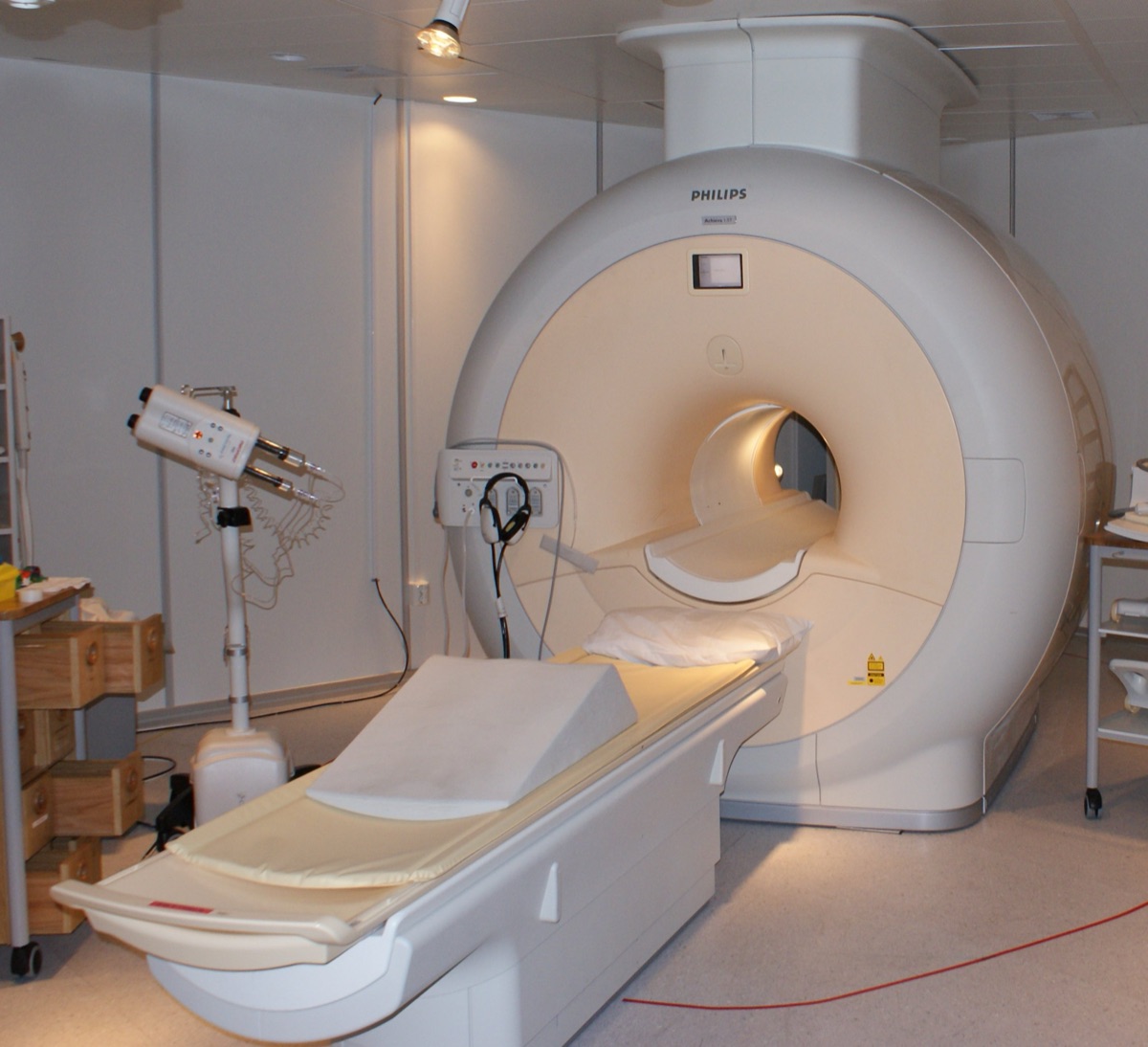
A magnetic resonance imaging scanner is a marvel of a magnet - but the part that actually makes the picture is a small radio antenna that whispers to the atoms in your body and then listens for the echo. A team at Berlin’s Max Delbrueck Center for Molecular Medicine decided to rebuild that antenna from first principles, weaving in metamaterials - engineered structures that bend electromagnetic waves in ways no natural material can. The result, published in Advanced Materials, is a transmitter-and-receiver that roughly doubles how well the scanner hears the body’s faint signals, and produces sharper images of some of the hardest regions to capture - the eye, the optic nerve, and the brain’s visual cortex - in less time. And it does all of this on the machines hospitals already own.
- What: a redesigned MRI radiofrequency antenna built from metamaterials (split-ring resonators)
- Receive sensitivity: up 94-132% - it roughly doubled how well the antenna detects the returning signal
- Transmit efficiency: up 14-20%; transmit-field strength up 13-21%
- In the living eye: measured signal up 25-51%
- Payoff: sharper images of the eye, optic nerve and visual cortex, in less scan time
- Best part: it slots into existing 7-tesla scanners - upgrade the antenna, not the magnet
- Published: Advanced Materials (DOI 10.1002/adma.202517760); Max Delbrueck Center + Rostock University Medical Center
1. The Overlooked Part of an MRI Machine
When people picture an MRI, they picture the giant doughnut - the superconducting magnet that lines up the protons in your body’s water molecules. But the image itself is made by a second, far humbler component: the radiofrequency (RF) antenna, also called a coil. It sends a pulse of radio waves to gently tip those protons, then acts as a microphone to pick up the whisper-faint signal they give off as they settle back. The quality of your scan depends enormously on how efficiently that antenna can transmit the pulse and receive the echo.
That is exactly the piece the Berlin team rebuilt. Crucially, they left the multimillion-dollar magnet untouched. Every gain here comes from a smarter antenna - which is why it can, in principle, be added to scanners that are already installed.
2. What a Metamaterial Actually Does Here
A metamaterial gets its properties not from its chemistry but from its structure - a repeating pattern of tiny engineered elements, each far smaller than the wavelength it manipulates. Arrange them cleverly and you can steer, focus or slow electromagnetic waves in ways ordinary materials cannot.
Here the building block is the split-ring resonator: a small conductive ring with a gap, which resonates with the radio field and reshapes it. The researchers arranged a grid of these unit cells into an antenna that concentrates the RF field toward the target tissue instead of letting it spread out and weaken. More of the pulse reaches where it is needed, and more of the returning signal is captured - the electromagnetic equivalent of cupping your hands around your ears to hear a faint sound.
The study was done on an ultra-high-field 7-tesla scanner - the most powerful class of MRI now used on people (typical hospital machines are 1.5 or 3 tesla). Higher field means more raw signal and finer detail, but it also makes the radio physics trickier: the RF wavelength shrinks inside the body and the field becomes uneven. A better-designed antenna is precisely how you tame that - which is why 7T is the ideal proving ground for this idea.
3. The Numbers
Against a conventional antenna, the metamaterial design delivered large, measurable gains - on the bench and in living people:
| Measure | Improvement (7T study) |
|---|---|
| Receive sensitivity (hearing the echo) | +94% to +132% (roughly 2x) |
| Transmit efficiency (sending the pulse) | +14% to +20% |
| Transmit-field (B1+) strength | +13% to +21% |
| Live signal inside the human eye | +25% to +51% |
The single most striking figure is receive sensitivity: nearly doubling - and in places more than doubling - how well the antenna picks up the signal. In imaging, that extra sensitivity can be spent two ways: on a sharper picture at the same speed, or on the same picture faster (a shorter, more comfortable scan). Importantly, the eye gains were confirmed in vivo - in real volunteers, not just simulations.
4. Why the Eye and the Back of the Brain Are So Hard
The team aimed at two of the trickiest targets in the body. The eye and orbit - the eyeball, the extraocular muscles that move it, and the optic nerve - are small, delicate, and prone to motion, so getting a crisp image is a real challenge. The occipital lobe, the visual-processing hub at the very back of the head, sits deep and far from a surface antenna, where signal naturally fades.
These are not niche curiosities. Sharper, faster imaging of exactly these regions is valuable across ophthalmology and neurology - and demonstrating the gains here, where imaging is hardest, is a strong sign the approach can help elsewhere too. The researchers are already adapting the design for the heart and kidneys.
Senior author Prof. Thoralf Niendorf framed the payoff plainly: the work points toward “faster, clearer MRI scans that could benefit patients” across many areas of care. Lead author Nandita Saha captured the mindset behind it: the goal was “to rethink MRI hardware from the modern physics of antenna design.” Because the antenna shapes the radio field so precisely, the team notes it can even be tuned to protect sensitive areas - for instance, reducing unwanted heating around metal implants.
5. Why It Matters: A Smarter Machine, Not a Bigger One
The most appealing thing about this result is how practical it is. There is no new magnet, no new scanner room, no new multimillion-dollar purchase. The intelligence lives in the antenna - a component that can be swapped and refined. That is the kind of upgrade that can spread through existing hospitals rather than waiting a generation for new machines.
It also lands safely: the design kept tissue heating to a fraction of a degree and stayed comfortably within international RF safety limits, even while pushing more signal. Better images, shorter scans, and a gentler experience for the person in the tube - all from rethinking one overlooked part.
The Honest Caveats
- It was shown at 7 tesla. Most clinics run 1.5- or 3-tesla machines; adapting the antenna to those (and to routine clinical workflows) is the next engineering step.
- The in-body tests were small. The eye measurements confirm the gains in real people, but larger studies are needed to prove the clinical benefit - which is exactly what the team is now planning across several hospitals.
- This is a hardware advance, not a product. It is a demonstrated design principle, not yet an approved, shipping clinical coil.
Sources
- Saha, Niendorf et al., ‘Metamaterial Antennas Enhance MRI of the Eye and Occipital Brain,’ Advanced Materials (2026), DOI 10.1002/adma.202517760
- Max Delbrueck Center: New material boosts MRI image quality
- Physics World: Metamaterial antennas enhance MR images of the eye and brain
- ScienceDaily: New MRI breakthrough reveals the brain and eye like never before · SciTechDaily coverage
Curated by Jerry Cards - jerrycards.com. We research the week’s most consequential tech, science, and health news so you don’t have to. More at jerrycards.com/news.