
 العربية
 Español
 中文
中文 Deutsch
 Français
 Português
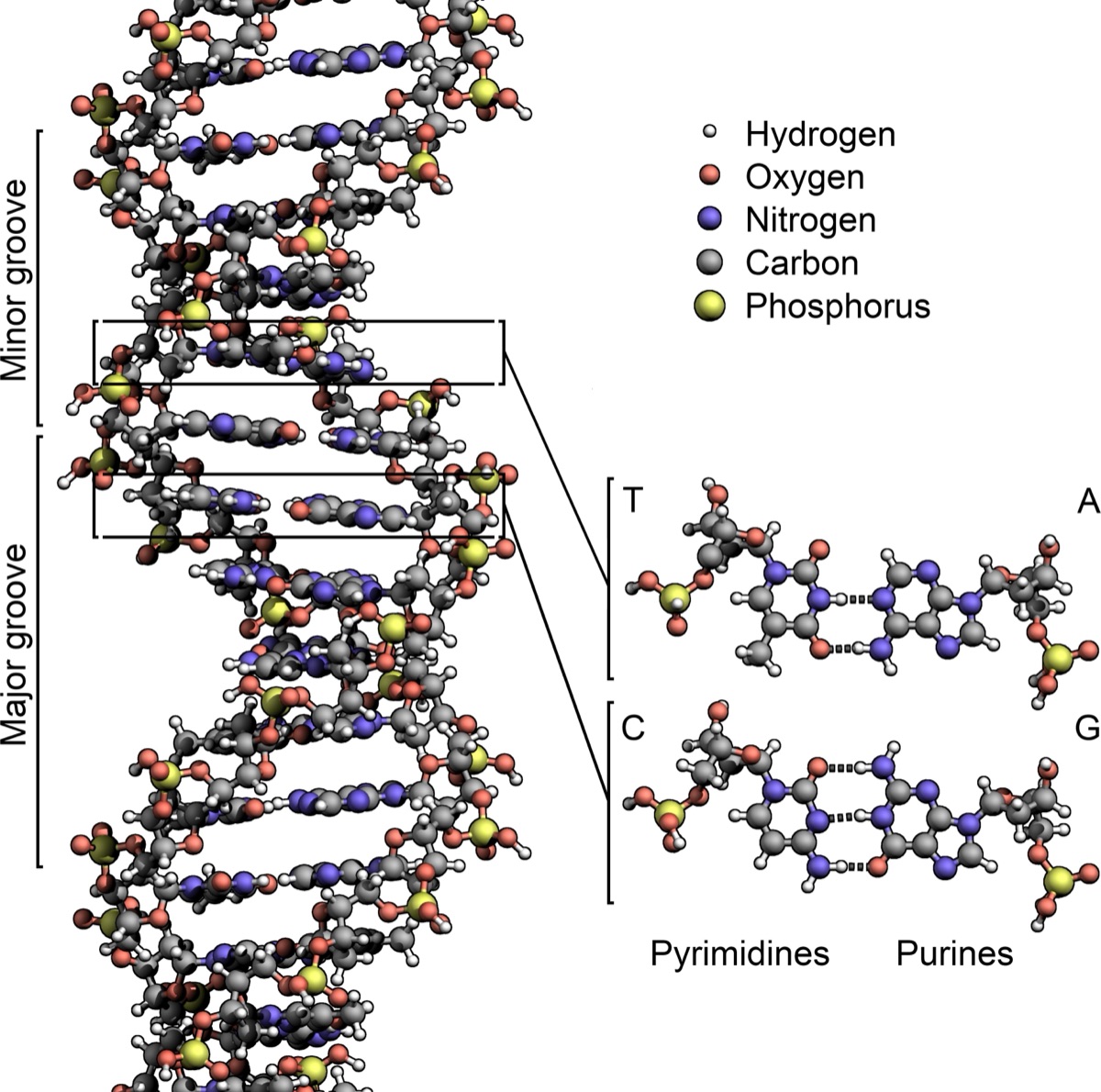
On April 25, 1953, a paper barely a page long appeared in the journal Nature - and quietly changed everything we know about living things. Its two young authors, James Watson and Francis Crick, proposed a structure for DNA, the molecule that carries heredity from one generation to the next. That structure - the double helix - did more than describe a molecule. Hidden inside its elegant shape was the answer to a question people had asked for millennia: how does life copy itself? It was the moment biology became molecular, and the modern sciences of genetics, genomics and biotechnology were born on that page.
This is a tribute to what Watson and Crick wrote, the people whose work made it possible, and how one short paper still shapes medicine, technology and our sense of who we are.
- Authors: J. D. Watson and F. H. C. Crick, working at the Cavendish Laboratory, University of Cambridge
- Title: “Molecular Structure of Nucleic Acids: A Structure for Deoxyribose Nucleic Acid”
- Published: Nature, vol. 171, pp. 737–738, 25 April 1953 (DOI 10.1038/171737a0)
- Length: about 800 words - roughly a single page, with one hand-drawn diagram
- The big idea: DNA is a double helix of two complementary strands; the base pairing (A–T, G–C) shows how the molecule can copy itself
- Built on: the X-ray images of Rosalind Franklin & Maurice Wilkins and the base-ratio rules of Erwin Chargaff
1. A question as old as life
By the early 1950s, biologists were fairly sure that DNA - deoxyribonucleic acid - was the stuff of genes. Experiments had shown it could carry hereditary information from one bacterium to another. But knowing DNA was the message only sharpened the mystery: what did the molecule look like, and how could a chemical possibly store a family resemblance and then copy it, letter-perfect, into every new cell? Nobody could answer that without knowing the shape.
Several groups were racing to find it. At the Cavendish Laboratory in Cambridge, Watson (a 24-year-old American biologist) and Crick (a 36-year-old physicist-turned-biologist) were building physical models out of cardboard and metal, trying to make the chemistry click into place. At King’s College London, Rosalind Franklin and Maurice Wilkins were aiming X-rays at DNA fibres to photograph the molecule directly. And in California, the great chemist Linus Pauling had just proposed a structure of his own - a triple helix that turned out to be wrong.
2. Two clues that cracked it open
Two pieces of evidence, from other people, were decisive.
The first was a photograph. Using X-ray crystallography, Franklin and her student Raymond Gosling had captured a now-famous image known as Photo 51 - a striking X-shaped pattern that is the unmistakable signature of a helix. It also encoded the molecule’s dimensions. When Watson saw the image, the helical shape - and its scale - snapped into focus.
The second was a rule about counting. The biochemist Erwin Chargaff had measured the four bases in DNA - adenine (A), thymine (T), guanine (G) and cytosine (C) - across many species and found a curious regularity: the amount of A always roughly equalled the amount of T, and G always roughly equalled C. Nobody knew why. Watson and Crick would explain it in a single stroke.
3. The insight: complementary base pairing
Here is the idea at the heart of the paper. Picture a ladder, then twist it into a gentle spiral. The two long rails are backbones of sugar and phosphate. The rungs are the bases, meeting in the middle - and this is the crucial part - they pair up in a fixed way: A always bonds to T, and G always bonds to C, each pair held together by weak hydrogen bonds (two in an A–T pair, three in a G–C pair). A and T fit together like matching puzzle pieces; so do G and C. No other combination fits the space.
That one constraint instantly explained Chargaff’s rule: if every A faces a T and every G faces a C, then of course the totals come out equal. But it did something far more profound. It meant the two strands are complementary - each is the negative image of the other. Know the order of letters on one strand, and the other is completely determined.
Each strand is a template for its partner. Split the ladder down the middle, and each half carries all the information needed to rebuild the whole.
4. The shape itself
The structure Watson and Crick described is beautiful in its regularity - two strands running in opposite directions (antiparallel), wound into a right-handed helix with the paired bases stacked neatly inside like the steps of a spiral staircase.
| Feature | Watson-Crick model |
|---|---|
| Overall shape | A right-handed double helix (two coiled strands) |
| Backbones | Sugar-phosphate chains on the outside, running in opposite directions |
| Rungs | Paired bases inside: A with T, G with C |
| Diameter | About 2 nanometres (20 ångströms) |
| One full turn | About 10 base pairs, rising ~3.4 nm (34 ångströms) |
Watson and Crick did not run a single new experiment for this paper. They reasoned their way to the answer by fitting the known chemistry and the X-ray measurements into a model that had to be internally consistent - and only one arrangement worked.
5. The most understated sentence in science
They knew exactly what they had. Rather than trumpet it, they wrote a closing line that has become legendary for its restraint:
“It has not escaped our notice that the specific pairing we have postulated immediately suggests a possible copying mechanism for the genetic material.”
Unpack that quiet sentence and you get the foundation of modern genetics. Because A pairs only with T and G only with C, a DNA molecule can unzip into two single strands, and each strand can act as a mould for a fresh complementary partner - producing two identical double helices where there was one. That is how genes are copied every time a cell divides, and how they are handed down from parent to child. (The mechanism, called semiconservative replication, was confirmed experimentally five years later.) In a single clause, Watson and Crick had linked a molecule’s shape to the deepest property of life: the ability to reproduce.
6. It took more than two people
The double helix is often told as a two-man triumph, but the discovery stood on a wider foundation, and a full tribute names everyone.
Rosalind Franklin produced the crucial X-ray evidence, including Photo 51 and precise measurements of the molecule’s geometry; her data were essential to getting the model right. She published her own supporting analysis in the very same issue of Nature. For decades her role was underplayed, but modern historians increasingly regard her as an equal contributor to the discovery rather than a bystander. Maurice Wilkins, also at King’s College London, shared in the X-ray work and, later, the Nobel Prize. And Erwin Chargaff’s base-ratio rules were the quiet clue that made the pairing click.
The 25 April 1953 issue of Nature carried three back-to-back papers on DNA: Watson and Crick’s model, a supporting X-ray paper by Wilkins, Stokes and Wilson, and one by Franklin and Gosling. Read together, the theory and the experimental evidence arrived on the same day.
What the double helix made possible
| Because we knew the structure... | ...we could build |
|---|---|
| Reading DNA | DNA sequencing and the Human Genome Project - the full readout of our genetic code (2003) |
| Copying DNA | PCR, the copy-and-amplify method behind diagnostics, research and DNA tests |
| Editing DNA | CRISPR gene editing and gene therapies for once-untreatable diseases |
| Writing with the code | mRNA vaccines, engineered proteins and modern biotechnology |
| Identifying with DNA | Genetic fingerprinting, ancestry testing and evolutionary biology |
Recognition, and a legacy
The story goes that on 28 February 1953, as the model came together, Crick walked into a Cambridge pub and announced that they had “found the secret of life.” He was not far off. In 1962, Watson, Crick and Wilkins shared the Nobel Prize in Physiology or Medicine for the discovery. Rosalind Franklin, whose images had been so pivotal, had died in 1958 - four years too early; the Nobel is never awarded posthumously. Today her contribution is widely honoured alongside theirs.
The date itself has become a small monument. April 25 - the day the paper appeared, and also the day the human genome was declared complete half a century later, in 2003 - is now marked around the world as DNA Day.
Why it still matters
Every time a doctor orders a genetic test, a lab reads a cancer’s mutations, a vaccine is designed from a virus’s genetic sequence, or a person traces their ancestry from a cheek swab, they are using the insight that fit on that single page in 1953. The double helix turned heredity from a mystery into an engineering problem we could finally read, copy, and eventually rewrite.
What makes the story endure is its shape - simple, symmetrical, and true. Two strands, four letters, one rule for pairing them, and out of that spare vocabulary comes the endless variety of life. Some papers report a result. A rare few hand us a new alphabet. Watson and Crick - with Franklin, Wilkins and Chargaff - handed us the one that every living thing is written in.
Sources & further reading
- Watson, J. D. & Crick, F. H. C. “Molecular Structure of Nucleic Acids: A Structure for Deoxyribose Nucleic Acid”, Nature 171, 737–738 (1953)
- Wikipedia: Nucleic acid double helix · Rosalind Franklin · Chargaff’s rules
- The Nobel Prize: Nobel Prize in Physiology or Medicine 1962 (Crick, Watson, Wilkins)
- U.S. National Library of Medicine, Profiles in Science: The Discovery of the Double Helix, 1951–1953
- Image: DNA double-helix structure diagram by Zephyris, Wikimedia Commons, CC BY-SA 3.0
Curated by Jerry Cards - jerrycards.com. Our 致敬 (tribute) series celebrates the landmark papers and discoveries that quietly built the modern world. More at jerrycards.com/news.